RF MICRONEEDLING AFTERCARE
Persistent Bumps After Needle RF (Potenza RF Microneedling, etc.)
How to distinguish expected swelling from dermatitis, infection, thermal injury, or persistent grid marks
Written by M.D. Bo Won Lee, Director of Springday Clinic Sinchon, Seoul.
Three-line overview
Small, evenly distributed bumps after RF microneedling are often caused by localized edema and inflammation.
Progressive pain, warmth, pus, blisters, dusky discoloration, or firm persistent papules require medical assessment.
Barrier care and diagnosis should come before massage, multiple topical medications, or additional energy-based treatments.
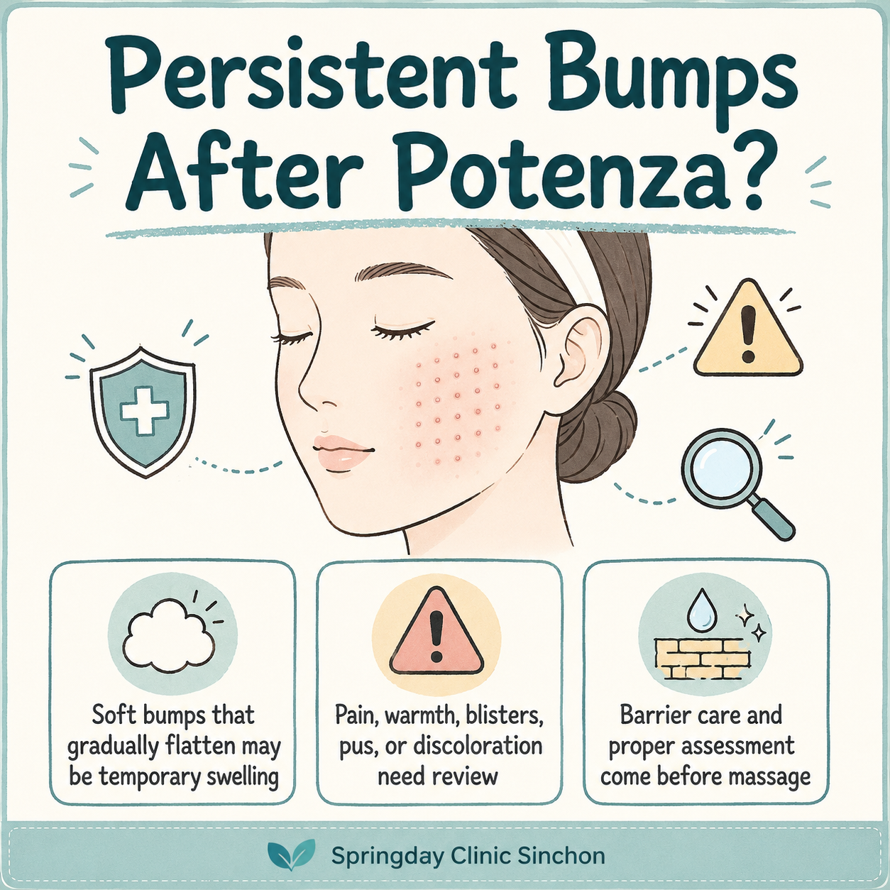
Soft, uniform elevations that become flatter and less red each day.
Increasing pain, heat, spreading redness, pustules, blisters, discoloration, or firm nodules.
Aggressive massage, picking, unprescribed medication mixtures, and premature repeat procedures.
Why do grid-like bumps appear?
Potenza is a radiofrequency microneedling platform. Microneedle electrodes penetrate the skin and deliver RF energy to create controlled electrothermal coagulation. The device can be used with different needle configurations, depths, RF modes, power levels, pulse durations, and treatment passes.
Because each needle creates a localized mechanical and thermal response, temporary swelling may reproduce the shape of the needle array. Patients often describe this appearance as embossing, stamping, or grid marks. These terms describe an appearance rather than a specific diagnosis.
Key point: The duration alone is not sufficient. Pain, temperature, color, firmness, surface damage, and the direction of change are equally important.
What is the expected recovery pattern?
Published reviews describe erythema, edema, dryness, pinpoint bleeding, and mild crusting as common short-term reactions. These responses often peak within the first 24 to 48 hours and gradually improve over approximately three to seven days. Results may vary according to the device settings, treatment site, skin thickness, previous procedures, and individual wound-healing response.
There is no universally accepted day on which persistent bumps automatically become a complication. As a practical clinical threshold, a lesion that has not improved after one week, or is becoming firmer or more symptomatic, deserves reassessment.
Five conditions that can look similar
1. Persistent localized edema
This is the most common explanation. The elevations are usually soft, relatively uniform, minimally tender, and progressively flatter. Thin areas, including the periorbital and upper cheek regions, may make minor swelling appear more prominent.
2. Irritant or allergic contact dermatitis
The temporarily disrupted barrier may react to anesthetic cream, antiseptics, adhesives, post-procedure masks, fragranced products, or active skin care. Itching, diffuse redness, burning, dryness, and scaling suggest dermatitis rather than simple edema.
3. Folliculitis or infection
Pustules, expanding redness, increasing warmth, tenderness, drainage, or yellow crusting require assessment for bacterial infection. Grouped blisters near the lips or nose may indicate herpes simplex reactivation. Treatment should be selected according to the suspected organism and clinical severity.
4. Excessive thermal injury
Blistering, erosions, sharply demarcated crusts, persistent pain, or pale, dusky, purple, or grey discoloration are not typical features of uncomplicated swelling. Thermal injury may result from a combination of energy, pulse duration, treatment depth, repeated passes, overlap, tissue thickness, and anatomical location.
5. Foreign-body or granulomatous inflammation
Rarely, firm papules or nodules may persist for weeks after microneedling, particularly when topical substances have been introduced through newly created channels. This cannot be determined without an in-person assessment. Culture, biopsy, or special staining may be necessary when infection and granulomatous inflammation are considered.
A mobile-friendly comparison
Soft, uniform, mildly red, minimally painful, and improving each day.
Itching, diffuse redness, burning, scaling, or worsening after a new product.
Increasing pain, warmth, spreading redness, pustules, blisters, erosions, or abnormal color.
What can be done at home?
- Simplify skin care. Pause retinoids, exfoliating acids, scrubs, strong vitamin C products, benzoyl peroxide, fragranced products, and alcohol-based toners.
- Cleanse gently. Use lukewarm water, a mild cleanser, and a simple moisturizer without rubbing the skin.
- Use brief cooling. A clean, cool pack wrapped in cloth may be applied for five to ten minutes. Do not place ice directly on the skin.
- Avoid heat and sweating. Temporarily avoid saunas, hot baths, intensive exercise, heavy alcohol intake, and prolonged sun exposure.
- Do not pick or massage. Mechanical pressure may worsen inflammation, bleeding, and post-inflammatory pigmentation.
- Document the course. Take photographs under consistent lighting to determine whether the bumps are actually improving.
Seek prompt medical review when
□ Pain or warmth is increasing rather than improving.
□ Redness is spreading beyond the treated pattern.
□ Pustules, drainage, yellow crusts, or grouped blisters appear.
□ The skin becomes pale, grey, purple, or dark red.
□ Bumps become distinctly firm or nodular.
□ Numbness, electric pain, weakness, or abnormal facial movement occurs.
□ There is no meaningful improvement after approximately one week.
□ A sharply visible grid remains unchanged for two weeks or longer.
How should a clinic assess persistent bumps?
A useful evaluation includes the tip type, needle insulation, treatment depth, monopolar or bipolar mode, power, pulse duration, number of passes, degree of overlap, anatomical site, and any substance applied or delivered immediately after the procedure.
The physical examination should document tenderness, warmth, blanching, pustules, vesicles, erosion, crusting, discoloration, firmness, sensory changes, and the direction of change. Bacterial culture, herpes testing, or biopsy is not required in every case, but may be appropriate when the clinical pattern is atypical.
Treatment principles
Barrier support, brief cooling, sun protection, and observation.
Remove the suspected trigger and consider a clinician-directed short anti-inflammatory regimen.
Use antimicrobial treatment only when clinically indicated, with culture when appropriate.
Prioritize wound care and follow-up for infection, pigmentation, texture change, and scarring.
Current evidence does not support a definitive conclusion that ultrasound, LED, laser, or another energy-based device reliably shortens persistent Potenza embossing. Additional energy should not be applied before infection, dermatitis, or thermal injury has been excluded.
Who may require closer follow-up?
Soft, non-tender, evenly distributed bumps with clear day-to-day improvement and no surface breakdown.
Progressive symptoms, immunosuppression, a history of abnormal scarring, active inflammatory skin disease, pustules, vesicles, erosions, or fixed firm papules.
Frequently asked questions
References
- Varelas EA, et al. Complications of radiofrequency microneedling: a comprehensive review. BMC Plastic and Reconstructive Surgery. 2026;2:21. DOI: 10.1186/s44452-026-00031-x.
- Kumar N, et al. Effectiveness of Radiofrequency Microneedling in the Treatment of Dermatological Conditions: A Systematic Review. Aesthetic Plastic Surgery. 2026. DOI: 10.1007/s00266-026-05834-y.
- Tan MG, et al. Radiofrequency Microneedling: A Comprehensive and Critical Review. Dermatologic Surgery. 2021;47(6):755-761. DOI: 10.1097/DSS.0000000000002972.
- Camacho-Hubbard I, et al. Safety Considerations and Complications With Radiofrequency Microneedling. Dermatologic Surgery. 2026. DOI: 10.1097/DSS.0000000000005251.
- U.S. Food and Drug Administration. Potential Risks with Certain Uses of Radiofrequency Microneedling. Safety Communication. 2025.
This article provides general medical information and does not replace an in-person diagnosis. Results may vary, and the correct management depends on examination findings, treatment records, and the direction of change over time.
#Potenza Aftercare, #RF Microneedling, #Potenza Bumps, #Grid Marks, #Microneedling Swelling, #Contact Dermatitis, #Folliculitis, #Thermal Injury, #Skin Barrier Repair, #Procedure Safety


