Redness, Pigmentation, and Enlarged Pores: Why Treatment Order Matters
CLINICAL SKIN TREATMENT GUIDE
Redness, Pigmentation, and Enlarged Pores: Why Treatment Order Matters
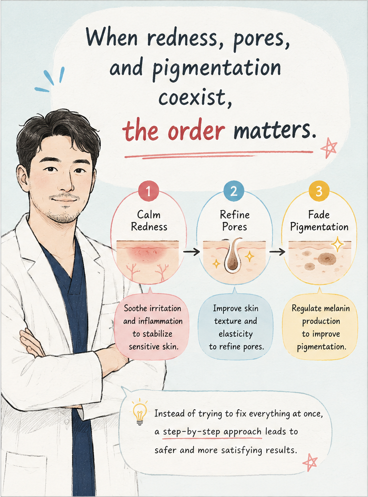
When redness, brown discoloration, and enlarged pores appear on the same face, simply combining three procedures is not always the most rational approach.
Written by M.D. Bo Won Lee, Director of Springday Clinic Sinchon, Seoul.
Clinical overview
There is no universally validated sequence that requires every patient to undergo redness treatment first, pigmentation treatment second, and pore treatment last. The order should be based on active inflammation, vascular findings, pigment type, skin tone, barrier function, and the mechanism making the pores appear enlarged.
REDNESS
Determine whether the redness is vascular, inflammatory, barrier-related, or transient.
PIGMENT
Distinguish melasma, lentigines, freckles, and post-inflammatory hyperpigmentation.
PORES
Assess sebum, comedones, follicular structure, dermal laxity, and acne scarring.
Three Visible Concerns Do Not Necessarily Mean Three Independent Problems
A patient may have persistent cheek redness, brown patches over the cheekbones, and visible pores around the nose at the same time. It is understandable to assume that one vascular procedure, one pigment laser, and one fractional treatment should simply be combined.
However, these findings exist within the same biological system. Vascular reactivity, inflammation, melanocyte activity, sebaceous function, the follicular opening, epidermal barrier integrity, and dermal support can influence one another.
The key question is not how many devices can be used, but which problem may interfere with the safety or effectiveness of the next treatment.
First, Identify What Is Causing the Redness
“Facial redness” is a visual description rather than a single diagnosis. Several different conditions may produce a red appearance.
Transient flushing
The face may become temporarily red after heat exposure, exercise, alcohol, spicy food, or emotional stress. This is not necessarily the same as persistent erythema or fixed telangiectasia.
Persistent erythema and visible vessels
Redness that remains visible at baseline, particularly when accompanied by fine facial vessels, may be considered for vascular-selective laser or intense pulsed light treatment after an appropriate assessment.
Inflammatory redness
Rosacea papules and pustules, acne, contact dermatitis, or seborrheic dermatitis may produce redness that cannot be explained by dilated vessels alone. Treating the underlying inflammation may be more important than immediately treating the visible red color.
Barrier-related sensitivity
Burning, stinging, scaling, and discomfort after cleansing may suggest that barrier dysfunction is contributing to the red appearance. Additional procedural irritation may be poorly tolerated until the skin is more stable.
Modern rosacea management is generally phenotype-based. Persistent erythema, telangiectasia, inflammatory lesions, and sensory symptoms are assessed individually rather than assuming that every patient with rosacea requires the same treatment. Laser and light-based therapies are options particularly for persistent erythema and telangiectasia, but they do not correct every cause of flushing.[1-3]
Brown Discoloration Must Be Classified Before It Is Treated
Brown facial lesions may represent freckles, solar lentigines, post-inflammatory hyperpigmentation, melasma, or another pigmented lesion. These conditions do not respond identically to treatment.
Discrete superficial pigmentation
Well-defined superficial freckles or lentigines may be considered for pigment-targeting lasers or IPL when the diagnosis and skin type are appropriate.
Post-inflammatory hyperpigmentation
PIH develops after inflammation or injury. If acne, dermatitis, or repeated irritation continues, new pigment may continue to appear even while existing pigment is being treated.
Procedures can also trigger additional PIH, particularly in patients with more melanized skin or a previous history of post-procedural pigmentation. A 2024 systematic review found that evidence for PIH treatment in skin of color remains heterogeneous, and laser-related worsening was reported in some patients.[4-5]
Melasma
Melasma is not equivalent to an isolated brown spot. It is a recurrent pigmentary disorder influenced by ultraviolet radiation, visible light, hormonal factors, inflammation, oxidative pathways, and, in some patients, vascular changes.
Laser and light procedures may be used as adjunctive treatments in selected cases, but aggressive or repetitive treatment can be followed by hyperpigmentation or hypopigmentation. Photoprotection and an appropriate long-term maintenance strategy remain fundamental.
Some melasma lesions demonstrate visible or subclinical vascular features. Small studies have evaluated combined vascular and pigment-targeting treatment in selected patients, but this does not establish a universal rule that vascular treatment must always come first.[8]
Enlarged Pores Are Not a Single Structural Problem
Pores do not open and close like doors. What patients commonly call enlarged pores is the visible appearance of the follicular opening and its surrounding structures.
Sebum-dominant pores
Excess sebum, keratin, blackheads, and comedones may make follicular openings appear darker and larger.
Laxity-related pores
Reduced dermal support may make pores appear vertically elongated.
Scar-related pores
Small atrophic acne scars may be mistaken for ordinary enlarged pores.
Fractional lasers, fractional picosecond systems, and radiofrequency microneedling have been studied for pore and texture improvement. A randomized split-face trial published in 2024 found both a 1064-nm picosecond laser with a microlens array and a 1565-nm non-ablative fractional laser to be viable options for enlarged pores. However, the trial was relatively small and does not identify one universally superior treatment.[6-7]
The realistic goal is usually to make pores less visible by treating the dominant mechanism, not to permanently erase every follicular opening.
Why Active Inflammation May Need to Be Addressed First
Inflammation and skin injury can activate melanogenesis and contribute to PIH. This is why acne, dermatitis, burns, and overly aggressive procedures may leave persistent brown or gray discoloration.
Fractional laser and radiofrequency microneedling treatments intentionally create controlled microscopic thermal or mechanical injury. This can be useful for remodeling scars and texture, but it also produces a temporary inflammatory response.
When the skin is already burning, scaling, inflamed, or highly reactive, stacking several energy-based treatments may increase recovery burden and narrow the safety margin. This does not mean that fractional treatments are inherently inappropriate. It means that the baseline condition of the skin and the cumulative treatment intensity matter.
A Practical Treatment-Planning Framework
Step 0 — Diagnose and classify
Determine the type of redness, the pigment diagnosis, the mechanism of pore enlargement, the patient’s skin tone, and previous treatment reactions.
Step 1 — Stabilize active inflammation and barrier dysfunction
Treat dermatitis, active inflammatory lesions, burning, or significant barrier impairment before adding unnecessary procedural stress.
Step 2 — Reassess persistent vascular redness
Consider vascular laser or IPL when persistent erythema or telangiectasia remains clinically relevant.
Step 3 — Treat pigment according to diagnosis
Separate melasma, lentigines, freckles, and PIH before selecting topical treatment, photoprotection, laser, or light-based treatment.
Step 4 — Address pores and texture
Select treatment based on sebum, comedones, laxity, follicular anatomy, and acne scarring.
This is a clinical framework, not an inflexible rule. A stable patient with superficial pigment and visible vessels may be suitable for a carefully selected combined light treatment. Another patient with active dermatitis or unstable melasma may benefit from a slower, staged approach.
When Same-Day Combination Treatment May Be Reasonable
Combination may be considered when
- the diagnoses are reasonably clear;
- the barrier is stable;
- the patient’s previous recovery pattern is known;
- the treatment intensity can be adjusted;
- cumulative thermal and inflammatory injury can be limited.
A staged plan may be safer when
- burning and stinging are present at baseline;
- dermatitis or inflammatory rosacea is active;
- melasma has recently worsened;
- the patient has a history of PIH;
- multiple high-intensity procedures would overlap.
Who May or May Not Be a Suitable Candidate?
A combined approach may be considered
For patients with stable skin, clearly identified superficial pigment and vascular findings, realistic expectations, and an acceptable recovery window.
A cautious staged approach may be preferable
For patients with active dermatitis, severe burning, recent sunburn, unstable melasma, ongoing inflammatory acne, or a strong history of post-procedural pigmentation.
Practical Checklist Before Treatment
□ Is the redness transient or persistent?
□ Are visible facial vessels present?
□ Is there burning, stinging, itching, or scaling?
□ Is acne, rosacea inflammation, or dermatitis active?
□ Is the brown discoloration melasma, lentigo, freckling, or PIH?
□ Has the patient previously developed PIH or a burn after treatment?
□ Are the pores related mainly to sebum, laxity, or scarring?
□ Will several treatments overlap on the same area?
□ Is sufficient recovery time available?
□ Is there a plan for post-treatment reassessment?
How I Assess Combined Skin Concerns at Springday Clinic Sinchon
Rather than automatically combining a redness package, a pigment package, and a pore package, I try to separate the following variables:
- whether the redness is active, inflammatory, or predominantly vascular;
- whether the pigment resembles melasma, discrete lentigines, or PIH;
- the patient’s baseline skin tone and history of post-treatment pigmentation;
- whether the pores are associated with sebum, laxity, or acne scarring;
- recent laser, peel, radiofrequency, or injectable treatment history;
- the patient’s highest-priority concern;
- the acceptable downtime and recovery schedule.
Frequently Asked Questions
References
[1] van Zuuren EJ, et al. Interventions for rosacea based on the phenotype approach. Br J Dermatol. 2019;181(1):65-79. DOI: 10.1111/bjd.17590. PMID: 30585305.
[2] Nguyen C, et al. Rosacea: Practical Guidance and Challenges for Clinical Management. Clin Cosmet Investig Dermatol. 2024;17:175-190. DOI: 10.2147/CCID.S391705. PMID: 38283794.
[3] Zhai Q, et al. Meta-Analysis of the Efficacy of Intense Pulsed Light and Pulsed Dye Laser in Rosacea. J Cosmet Dermatol. 2024. DOI: 10.1111/jocd.16549. PMID: 39240125.
[4] Ko D, et al. Disorders of hyperpigmentation. Part II. J Am Acad Dermatol. 2023;88(2):291-320. DOI: 10.1016/j.jaad.2021.12.065. PMID: 35158001.
[5] Mar K, et al. Treatment of Post-Inflammatory Hyperpigmentation in Skin of Colour. J Cutan Med Surg. 2024;28(5):473-480. DOI: 10.1177/12034754241265716. PMID: 39075672.
[6] Liu X, et al. Comparison of a 1064-nm picosecond MLA laser and 1565-nm non-ablative fractional laser for enlarged pores. Lasers Med Sci. 2024;39(1):80. DOI: 10.1007/s10103-024-04028-9. PMID: 38396012.
[7] Tan MG, et al. Radiofrequency Microneedling: A Comprehensive and Critical Review. Dermatol Surg. 2021;47(6):755-761. DOI: 10.1097/DSS.0000000000002972. PMID: 33577211.
[8] Kong SH, et al. Treatment of Melasma with Pulsed-Dye Laser and 1064-nm Q-Switched Nd:YAG Laser. Ann Dermatol. 2018;30(1):1-7. DOI: 10.5021/ad.2018.30.1.1.
Evidence limitation
No universal randomized protocol was identified that directly compares every possible treatment sequence for patients who simultaneously have facial redness, pigmentation, and enlarged pores. The framework above combines evidence for the individual conditions with practical clinical risk assessment.
Effective planning is not about treating everything at maximum intensity.
It is about selecting a sequence in which one procedure does not unnecessarily increase the risk or reduce the benefit of the next.
M.D. Bo Won Lee · Springday Clinic Sinchon · Seoul
#FacialRedness #Hyperpigmentation #EnlargedPores #RosaceaTreatment #PigmentationTreatment #LaserTreatment #SkinBarrier #SeoulSkinClinic #Sinchon #SpringdayClinicSinchon