RE2O NODULE ASSESSMENT
A Lump After Re2O or Re2O Fine: Why Diagnosis Must Come Before Dissolving Treatment
Written by M.D. Bo Won Lee, Director of Springday Clinic Sinchon, Seoul.
Three-line overview
Not every palpable lump after an injection is a granuloma.
Re2O products are particulate human acellular dermal matrix products, not standard hyaluronic acid fillers.
Observation, antibiotics, intralesional therapy, biopsy, and surgery apply to different clinical phenotypes.
Product
Particulate human acellular dermal matrix derived from skin tissue.
First decision
Determine whether the lesion is a deposit, infection, sterile inflammation, granuloma, or fibrosis.
Key caution
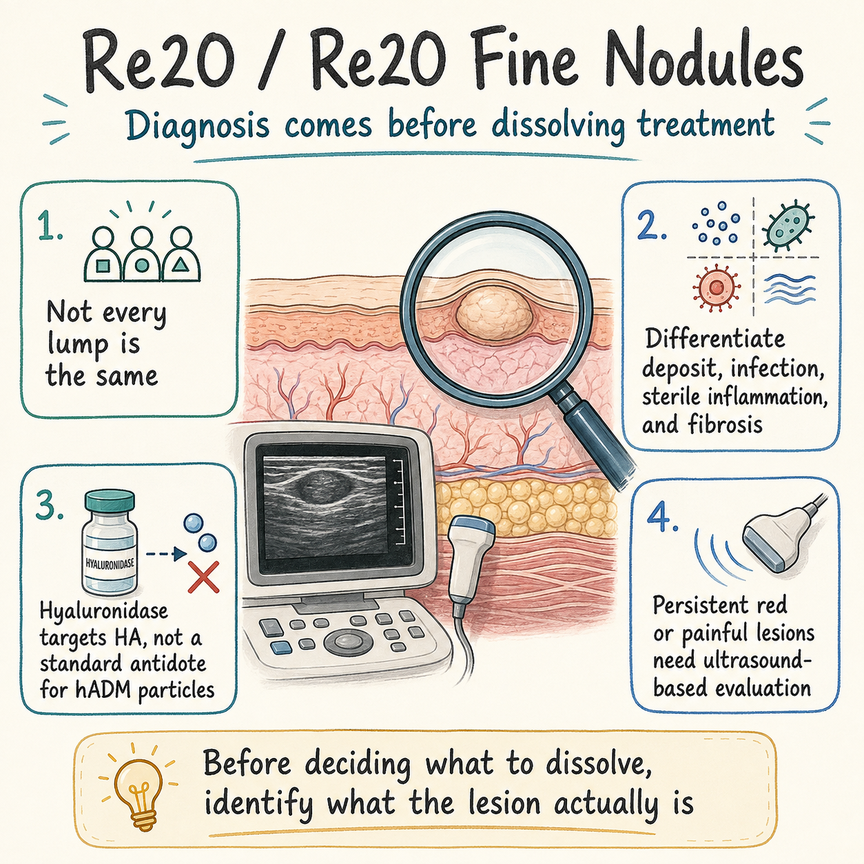
Hyaluronidase should not be presented as a direct antidote for the hADM particles.
The First Question Is Not “How Do We Dissolve It?”
A patient may notice a small bead, plaque, or firm lump after a Re2O procedure. The most useful first questions are when it appeared, whether it is shrinking or enlarging, and whether redness, heat, tenderness, drainage, or systemic symptoms are present.
Core principle: A lump is a physical finding, not a complete diagnosis.
What Are Re2O and Re2O Fine?
According to the manufacturer’s official product information, Re2O and Re2O Fine are particulate human acellular dermal matrix products derived from skin tissue. Re2O is listed as a 150 mg product, whereas Re2O Fine is listed as a 50 mg product with a finer particle profile.
Re2O
150 mg
Particulate acellular dermal matrix
Re2O Fine
50 mg
Finer particulate acellular dermal matrix
A smaller particle profile does not prove that nodule formation cannot occur. No direct comparative nodule-incidence study between the two products was identified. Results may vary with the anatomical site, skin thickness, injection plane, local volume, distribution, prior injectables, infection, and individual tissue response.
Why Hyaluronidase Is Not a Universal Solution
Hyaluronidase enzymatically degrades hyaluronic acid. Re2O products are not standard cross-linked HA fillers, so hyaluronidase should not be described as an agent that directly dissolves their hADM particles.
Hyaluronidase may still be relevant when a previous HA filler is present in the same area or when ultrasound identifies a separate HA deposit. In that setting, it targets the HA component rather than the dermal-matrix particles.
Four Clinical Patterns to Consider
1. Early Non-Inflammatory Deposit or Edema
A lump that appears immediately after treatment and gradually becomes smaller or softer, without redness, warmth, or increasing pain, may represent edema, bruising, superficial deposition, or localized clustering of material.
2. Infection or Abscess
Progressive erythema, warmth, tenderness, rapid swelling, fluctuance, drainage, fever, or spreading inflammation requires prompt assessment. Ultrasound, aspiration, culture, antibiotics, and drainage may be required depending on the findings.
3. Sterile Inflammatory Nodule or Foreign-Body Granuloma
A delayed firm lesion, recurrent swelling, or multiple inflammatory nodules without a confirmed infection may represent a sterile inflammatory or foreign-body response. Intralesional corticosteroid, corticosteroid combined with 5-fluorouracil, biopsy, or other interventions may be considered after infection has been reasonably excluded.
Evidence for these treatments comes mainly from other injectable materials. Current evidence does not support a definitive Re2O-specific dose or treatment interval.
4. Fibrotic or Encapsulated Lesion
A long-standing, fixed, nonfluctuant lesion with little active inflammation may be predominantly fibrotic. Repeated blind injections and aggressive manipulation may provide limited benefit and may increase tissue trauma.
When Ultrasound Adds Value
High-frequency ultrasound can help localize the lesion, assess fluid collections, identify prior filler deposits, characterize fibrosis, and guide targeted treatment. Doppler imaging may help evaluate inflammatory vascularity and the relationship to adjacent vessels.
Ultrasound cannot replace histopathology in every atypical case, but it can reduce guesswork before intralesional treatment.
Comparison Cards
Observation May Be Reasonable
Present from the early post-treatment period
Becoming smaller or softer
No progressive erythema or warmth
Minimal or no pain
Prompt Medical Review
Increasing size or tenderness
Redness, heat, or drainage
Fluctuance or rapid swelling
Fever or spreading inflammation
Further Diagnostic Workup
Delayed onset after an initially quiet period
Multiple or recurrent nodules
Very firm or fixed lesion
Poor response to initial management
A Stepwise Management Framework
Step 1: Document and Reassess
Record the exact product, treatment date, anatomical site, prior injectables, onset of the lump, and interval change. Standardized photographs and measurements are preferable to relying on memory.
Step 2: Exclude Urgent Conditions
Progressive pain, skin-color change, pustular drainage, fluctuance, systemic illness, or visual symptoms require prompt evaluation for infection, vascular injury, or another urgent complication.
Step 3: Manage Suspected Infection
When feasible, image and aspirate a fluid collection before antibiotics. Culture-directed antimicrobial treatment is preferred when a specimen is available. An abscess may require drainage. Intralesional corticosteroid should generally be deferred while clinically meaningful infection remains possible.
Step 4: Consider Intralesional Treatment for Sterile Inflammatory Disease
After infection has been reasonably excluded, intralesional corticosteroid may be considered. A 5-fluorouracil combination is used in some resistant granulomatous filler reactions, but this is not a product-specific Re2O protocol.
Step 5: Biopsy or Excision for Atypical or Refractory Lesions
Biopsy may be appropriate for progressive, ulcerated, draining, atypical, or treatment-resistant lesions. Surgical excision may be necessary for a localized refractory mass, but potential scarring, contour change, nerve or vascular injury, and incomplete removal should be discussed.
Treatments That Should Not Be Used Automatically
- Repeated hyaluronidase without evidence of an HA component
- Aggressive massage of a painful, red, or warm lesion
- Intralesional steroid before meaningful infection has been assessed
- Energy-based treatment presented as a proven method of dissolving hADM particles
- Additional filler or booster injection into an unresolved lesion
Checklist Before Treatment
□ Exact product and treatment date
□ Lot information if available
□ Onset and interval change
□ Previous HA or biostimulatory injectables
□ Redness, warmth, tenderness, drainage, or fever
□ Autoimmune disease or immunosuppressive medication
□ Ultrasound, culture, or biopsy requirement
□ Risks of atrophy, scarring, and contour change
Suitable and Less Suitable Candidates for Observation
Observation May Be Suitable
The lesion is clearly improving
No inflammatory or systemic symptoms
No concerning skin-color change
Reliable follow-up is available
Observation Alone Is Less Suitable
The lesion is enlarging or becoming painful
Redness, warmth, drainage, or fever
Delayed multiple nodules
Immunosuppression or recurrent infection
Frequently Asked Questions
References
- L&C Bio. Official product information for Re2O, particulate human acellular dermal matrix, 150 mg.
- L&C Bio. Official product information for Re2O Fine, finer particulate human acellular dermal matrix, 50 mg.
- Chiang J, Liao YH. Mapping Filler Nodules. Dermatol Surg. 2026. DOI: 10.1097/DSS.0000000000004963. PMID: 41677155.
- WFUMB Position Paper: Consensus on Best Practice in Aesthetic Dermatologic Ultrasound. DOI: 10.1016/j.ultrasmedbio.2025.07.003. PMID: 40866164.
- Mlosek RK, et al. High-frequency ultrasonography for palpable nodules after dermal fillers. DOI: 10.15557/JoU.2020.0044. PMID: 33500791.
- Kroumpouzos G, Treacy P. Hyaluronidase for Dermal Filler Complications. DOI: 10.2196/50403. PMID: 38231537.
- Flores Rodríguez JC, et al. Management of PLLA Nodules. DOI: 10.7759/cureus.110742. PMID: 42438624.
- Sivam S, et al. Giant PMMA Foreign Body Granulomas with Imaging. DOI: 10.4103/JCAS.JCAS_194_20. PMID: 38189071.
Results may vary. Evidence remains limited for product-specific Re2O nodule management. A persistent or inflammatory lesion requires a diagnosis-based plan rather than an automatic dissolving injection.